

We’re here to help, in whatever way we can
New Patient Form
Request a Refill
Preparing your animal for surgery
We know that surgery day can be stressful for pets and their owners. We’ll do everything we can to ease your fears, walking you through the process and answering any questions you might have. Let’s get started so that your pet can be well on their way to a healthy recovery! We work together with pet owners and their regular veterinarian every step of the way, from the first consultation through the healing process. Thank you for allowing us to care for your beloved companion.
Conditions and Procedures
Jump to: TTA Surgery, Animal Reproductive Status, Urinary Surgery, Upper Airway Conditions, TPLO Surgery, Minimally Invasive Surgery, Elbow Disease, Hip Dysplasia, Media Patella Luxation
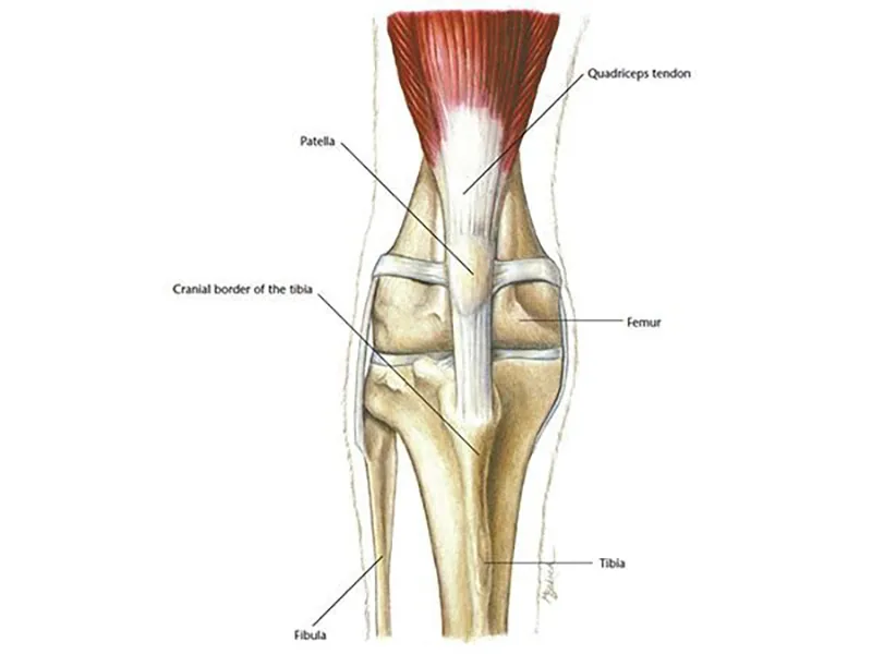
TTA surgery for cruciate ligament rupture
The cranial (or anterior) cruciate ligament in pets is located in the knee. A rupture of this ligament is the most common cause of rear leg lameness and can be quite severe due to the slide of the shin bone when the pet is moving. Tibial tuberosity advancement (TTA) surgery is designed to change the angle of the shin bone coming into the knee to stabilize the joint. The surgery does not directly repair the ruptured ligament, but it can offer major improvements in terms of walking and decreases in lameness.
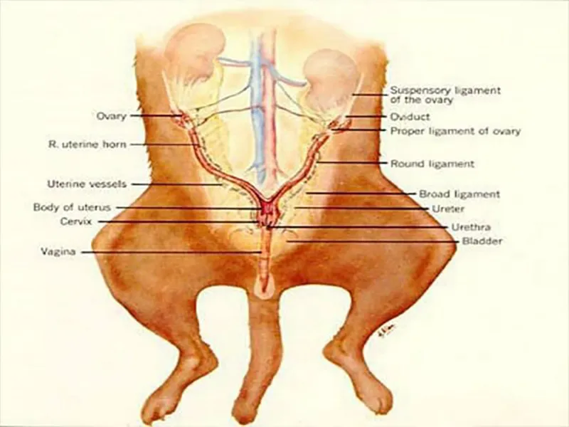
Ways to alter an animal’s reproductive status
At Lenity, we understand that managing an animal’s reproductive status is a crucial aspect of responsible pet ownership and animal welfare. There are various reasons why you might need to alter an animal’s reproductive status, such as controlling overpopulation, improving health, or addressing behavioral issues. In this guide, we explore the different methods available for altering an animal’s reproductive status, along with their benefits and considerations.
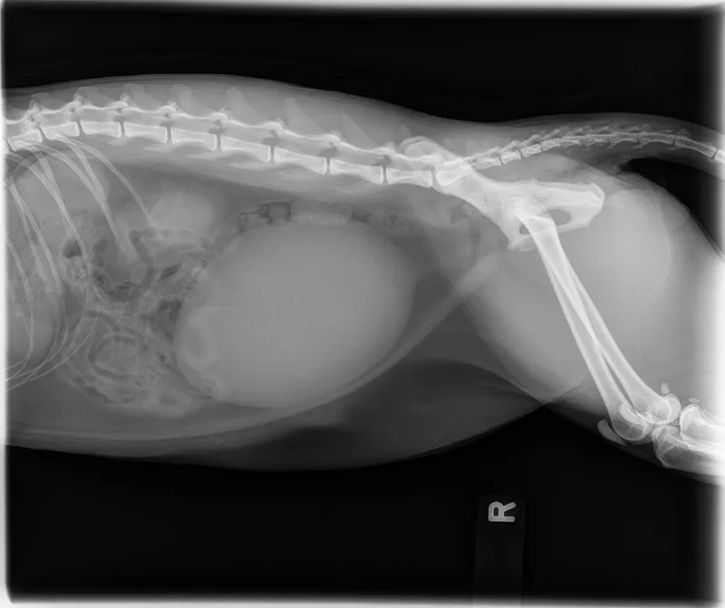
Urinary Surgery
At Lenity, we understand that urinary issues in pets can be a cause for concern and require prompt attention. Urinary surgery is a specialized field of veterinary medicine that focuses on diagnosing and treating various conditions affecting the urinary tract of animals. In this guide, we provide an overview of common urinary conditions, the surgical procedures involved, and how our experienced team at Lenity can help ensure the well-being of your beloved pet.
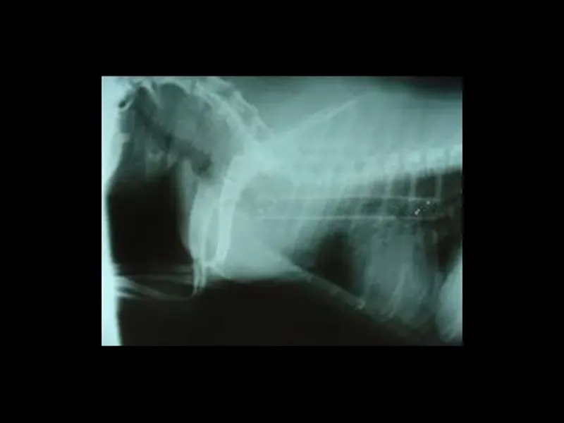
Upper Airway Conditions
Understanding Upper Airway Conditions
The upper airway of your pet includes the nasal passages, throat, and windpipe (trachea). Conditions affecting this area can lead to respiratory issues, impacting your pet’s ability to breathe comfortably.
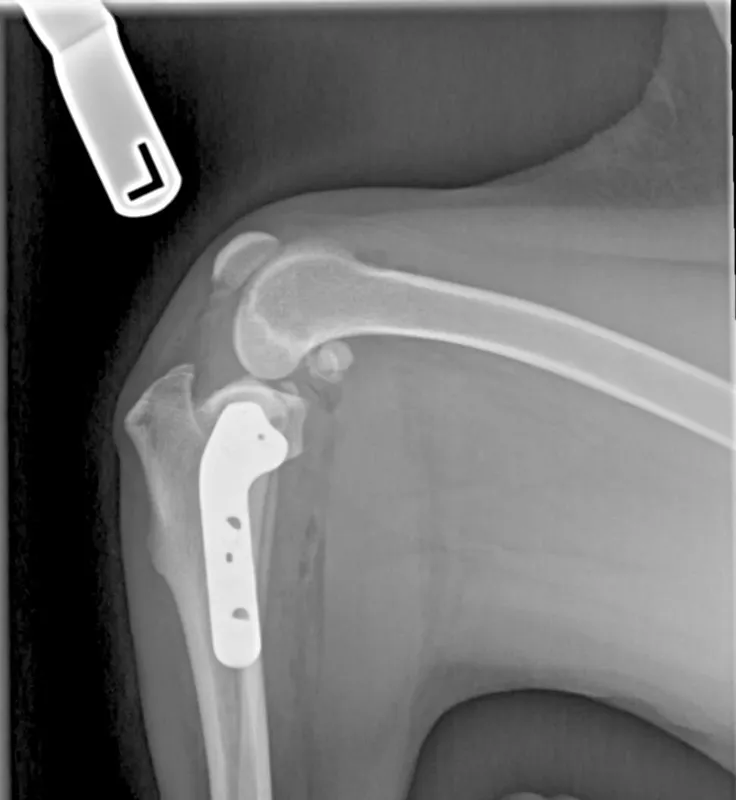
TPLO surgery for cruciate ligament injury
What is TPLO Surgery?
TPLO surgery is a specialized orthopedic procedure designed to treat cranial cruciate ligament (CCL) injuries in dogs. The cranial cruciate ligament is essential for stabilizing the knee joint, and when it is damaged, it can lead to pain, lameness, and joint instability. TPLO surgery involves restructuring the geometry of the knee joint to provide stability without relying on the function of the damaged ligament.
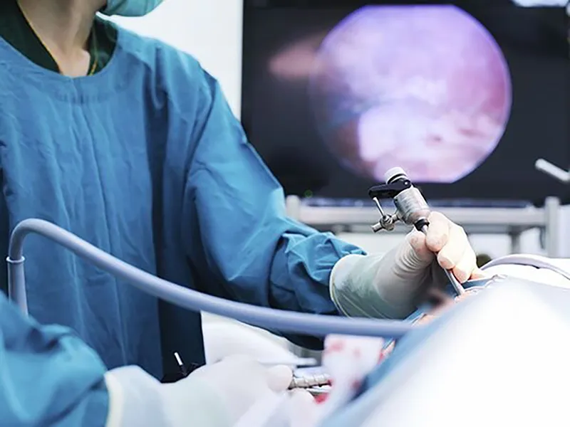
Minimally Invasive Surgery
What is Minimally Invasive Surgery (MIS)?
Minimally Invasive Surgery, often referred to as laparoscopic or endoscopic surgery, is a modern surgical technique that utilizes specialized instruments and cameras to perform procedures through tiny incisions or natural body openings. This approach minimizes trauma to surrounding tissues and organs, resulting in less pain, reduced scarring, and a shorter recovery period for your pet.
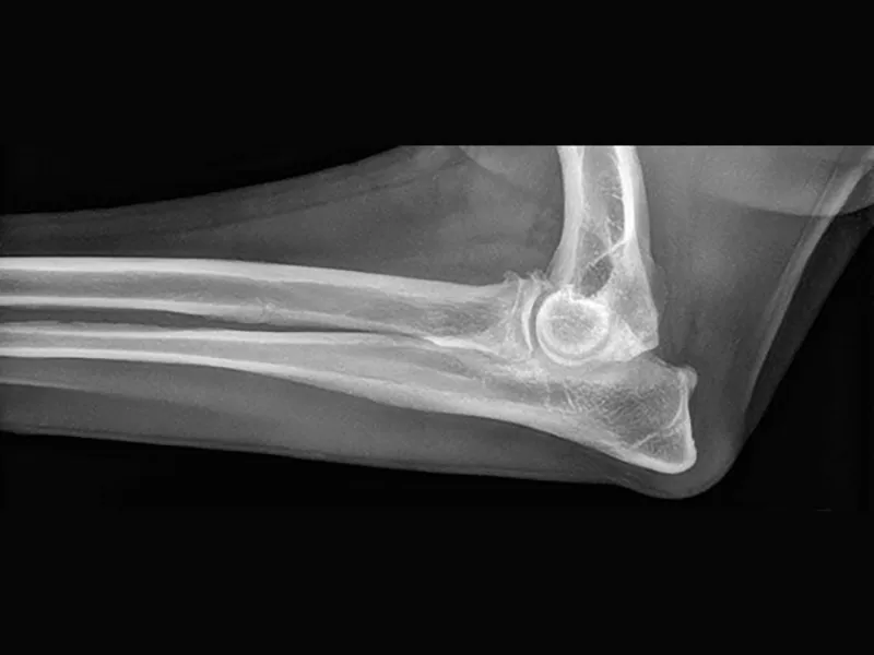
Elbow Disease
What is Elbow Disease?
Elbow disease, or elbow dysplasia, is a collective term for a group of developmental orthopedic conditions that affect the elbow joint in dogs, primarily in larger breeds. These conditions often involve abnormal growth and development of the bones and cartilage within the joint, leading to joint instability, pain, and arthritis.
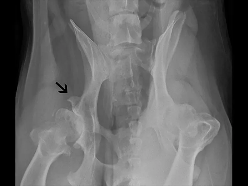
Hip Dysplasia
What is Hip Dysplasia?
Hip dysplasia is a genetic condition in which the hip joint does not develop properly. It occurs when the hip joint’s ball and socket do not fit snugly together, leading to instability and eventual damage to the joint. Over time, this can result in painful arthritis and reduced mobility.
Medial Patella Luxation
What is Medial Patella Luxation (MPL)?
Medial Patella Luxation is a common orthopedic condition in which the patella (kneecap) shifts out of its normal position, usually towards the inside of the knee joint. This misalignment can cause varying degrees of lameness and discomfort.